

USMLE Step 1 Anatomy Quiz 1 – 35 High-Yield Practice Questions + Answers (2026 Update)
1. A 36-year-old woman is hospitalized for treatment of a stomach ulcer that has been getting progressively worse over several months. Radiographic studies reveal the site of involvement to be along the greater curvature, approximately 4 cm away from the pyloric sphincter. That night, the ulcer perforates, and there is considerable intra-abdominal bleeding. Surgery reveals that the ulcer has eroded through the stomach wall and has damaged the artery supplying the involved region of the stomach. Which artery was likely involved?
- A. Left gastric
- B. Left gastroepiploic
- C. Right gastric
- D. Right gastroepiploic
- E. Short gastric
The left gastric artery (choice A), off the celiac trunk, supplies the left half of the lesser curvature of the stomach.
The left gastroepiploic artery (choice B), off the splenic artery, supplies the left half of the greater curvature of the stomach. Although it anastomoses with the right gastroepiploic artery, it is unlikely that this artery would be directly damaged by ulceration of the stomach near the pyloric sphincter.
The right gastric artery (choice C), off the proper hepatic artery, supplies the right half of the lesser curvature of the stomach.
The short gastric artery (choice E), actually one of several (4-5) short gastric arteries, off the splenic artery (occasionally the left gastroepiploic), supplies the fundus of the stomach, which is most distant from the pylorus.
2. A previously normal 56-year-old woman comes to the emergency department because of a “flu-like” illness. She complains of nausea and vomiting, unilateral tingling in the leg, and a headache involving the eye and forehead. She is alert and fully oriented. Motor, sensory, gait, and coordination examinations are normal. Cranial nerve examination is normal, aside from the visual field disorder indicated in the plot below.
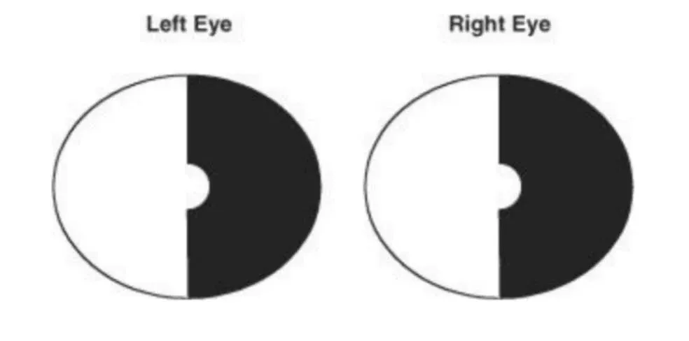
The neurologic examination suggests an occlusion of which of the following vessels?
- A. Left middle cerebral artery
- B. Left posterior cerebral artery
- C. Right middle cerebral artery
- D. Right posterior cerebral artery
- E. Posterior communicating artery
The middle cerebral artery (choices A and C) is the largest cerebral artery and supplies blood to most of the lateral surface of the hemisphere. However, it does not extend to the visual cortex.
The posterior communicating artery (choice E) is part of the circle of Willis and does not supply the cerebral cortex
3. A 49-year-old Vietnamese man is diagnosed with tuberculosis. On physical examination, large flocculent masses are noted over the lateral lumbar back, and a similar mass is located in the ipsilateral groin. This pattern of involvement strongly suggests an abscess tracking along which of the following muscles?
- A. Adductor longus
- B. Gluteus maximus
- C. Gluteus minimus
- D. Piriformis
- E. Psoas major
The gluteus maximus (choice B), gluteus minimus (choice C), and piriformis (choice D) are muscles of the buttock with no relationship to the groin.
4. A resident physician is performing a pelvic examination on a young woman. The fingers of one of her hands are in the patient’s vagina, palpating the cervix. The other hand is pressing on the abdomen. With the palm of this hand, the physician feels a bony structure in the lower midline. This structure is most likely the
- A. соссух
- B. ilium
- C. ischium
- D. pubis
- E. sacrum
The coccyx (choice A) is the caudal terminus of the vertebral column, generally formed by the fusion of four rudimentary vertebral bodies. It is also called the tailbone. The ilium (choice B) is one of the two “wings” that form the lateral sides of the pelvic cavity and support the abdominal contents.
The ischium (choice C) is the portion of the pelvis on which a person sits. The sacrum (choice E) is the triangular bone situated just beneath the lumbar vertebrae.
5. A 24-year-old man is stabbed in the neck. Neurologic examination reveals left-sided hemiparesis. There is complete loss of discriminatory joint position and vibration sensation below C8 on the left side. On the right side, there is loss of pain and temperature sensation below C8. It can be expected that an MRI of the cervical spinal cord will show which of the following findings?
- A. A complete transection of the spinal cord
- B. A hemisection of the left side of the cord
- C. A lesion of the dorsal columns of the cord on the left side
- D. A lesion of the lateral funiculus on the left side
- E. Damage to cervical dorsal roots on left side
The patient has the classic pattern of Brown-Séquard syndrome, caused by a hemisection of the spinal cord:
Ipsilateral (left) loss of motor function → due to damage of the left corticospinal tract.
Ipsilateral loss of vibration and proprioception → due to damage of the left dorsal columns (which ascend ipsilaterally).
Contralateral (right) loss of pain and temperature → due to damage of the left spinothalamic tract, which carries fibers that cross 1–2 segments above.
This triad matches a left hemisection of the cervical spinal cord, making B the correct answer.
6. A 2-year-old boy is brought to the pediatrician by his mother because he has had several episodes of rectal bleeding. Evaluation with a technetium-99m perfusion scan reveals a 3-cm ileal outpouching located 60 cm from the ileocecal valve. This structure most likely contains which of the following types of ectopic tissue?
- A. Duodenal
- B. Esophageal
- C. Gastric
- D. Hepatic
- E. Jejunal
Something else to keep in mind: A favorite question attendings ask on the wards is the rule of 2s associated with Meckel diverticulum: it occurs in about 2% of children, occurs within approximately 2 feet of the ileocecal valve, contains 2 types of ectopic mucosa (gastric and pancreatic), and its symptoms usually occur by age 2.
None of the other answer choices has a relationship to Meckel diverticulum.
7. A 34-year-old man is examined in the prison infirmary after sustaining a superficial stab wound to the superolateral aspect of the thoracic wall at the level of the third rib. There is little bleeding and no difficulty breathing; however, the medial border of the scapula on the injured side pulls away from the body wall when the arm is raised. In addition, the arm cannot be abducted above the horizontal. Which of the following muscles is paralyzed?
- A. Levator scapulae
- B. Pectoralis minor
- C. Rhomboid major
- D. Serratus anterior
- E. Supraspinatus
The levator scapulae (choice A) elevates the scapula. It is not involved in holding the scapula against the body wall. Its innervation, derived from the cervical plexus (C3, C4), is not damaged by injury to the superolateral thoracic wall.
Although the pectoralis minor (choice B) has a scapular attachment (to the coracoid process), it is not involved in holding the scapula against the body wall. Its innervation, the medial pectoral nerve, is not likely to be damaged by injury to the superolateral thoracic wall.
The rhomboid major (choice C) primarily retracts the scapula. It does not have an important role in holding the scapula against the body wall. Its innervation, the dorsal scapular nerve, is not easily damaged by injury to the superolateral thoracic wall. Although the supraspinatus (choice E) plays a role in arm abduction, it is essential to the initiation of this movement (the first 20–30 degrees) before there is significant rotation of the scapula. It is not involved in holding the scapula against the body wall (although it is important in holding the head of the humerus against the glenoid fossa). Its innervation, the suprascapular nerve, would not be damaged by injury to the superolateral thoracic wall.
8. A histologist is examining cells arrested at various stages of oogenesis. He discovers a follicle within the stroma of the ovary that has developed an antrum. This follicle would be classified as
- A. Graafian
- B. primary
- C. primordial
- D. secondary
The primary follicles (choice B) are relatively small and spherical, with a central oocyte and one or more layers of cuboidal-like follicular cells. The primary oocytes, which are arrested in prophase of their first meiotic division, are contained within the primordial follicles (choice C) as inactive reserve follicles. Recall the sequence of oogenesis:
Primordial follicle (contain primary oocytes) ⭢ Primary follicle ⭢ Secondary follicle ⭢ Graafian follicle
9. A 47-year-old man presents to his neurologist with an unsteady, broad-based gait and slow, slurred speech. Neurologic examination reveals dysdiadochokinesia, intention tremor, hypotonia, and nystagmus. The patient’s lesion is in a brain structure that derives from which of the following embryonic structures?
- A. Diencephalon
- B. Mesencephalon
- C. Metencephalon
- D. Myelencephalon
- E. Telencephalon
During the fourth week of embryonic development, the anterior end of the neural tube develops three vesicles: the prosencephalon (forebrain), the mesencephalon (midbrain), and the rhombencephalon (hindbrain). By the sixth week, 5 vesicles (listed in the answer options) have developed. The rhombencephalon has now developed into the metencephalon and myelencephalon. The cerebellum and pons derive from the metencephalon.
The diencephalon (choice A), which is derived from the prosencephalon, develops into the thalamus, hypothalamus, epithalamus, subthalamus (everything with the word “thalamus”), posterior lobe of the pituitary, and neural retina.
The mesencephalon (choice B), or midbrain, is the only brain vesicle that does not produce a secondary vesicle; it remains the mesencephalon. The myelencephalon (choice D), which is derived from the rhombencephalon, develops into the medulla oblongata.
The telencephalon (choice E), which is derived from the prosencephalon, develops into the cerebral hemispheres (cerebral cortex, basal ganglia, and deep white matter).
10. A 47-year-old woman with a history of rheumatic fever is examined by her physician. Physical examination is significant for a low-pitched, rumbling, diastolic murmur preceded by an opening snap. The affected valve can be best evaluated by auscultation at which of the following locations?
- A. Left second intercostal space
- B. Left fifth intercostal space
- C. Left lower sternal body border
- D. Right second intercostal space
- E. Right fifth intercostal space
The pulmonary valve is most audible over the left second intercostal space (choice A).
Pulmonic stenosis is associated with tetralogy of Fallot, a common cyanotic congenital heart disease.
The tricuspid valve is most audible over the left lower sternal border (choice C). This valve is primarily affected in IV drug abusers with endocarditis.
The aortic valve is most audible over the right second intercostal space (choice D).
Aortic stenosis is associated with angina, syncope, and congestive heart failure. A systolic murmur that radiates to the carotids is heard on exam, along with a systolic ejection click.
No particular structure is best heard over the right fifth intercostal space (choice E).
11. A newborn girl is noted to have abnormal accommodation of the lens. Further evaluation reveals an abnormal production of aqueous humor. A malformation of the structure responsible for these functions that is continuous posteriorly with the choroid might be due to abnormal development of
- A. axons of retinal ganglion cells
- B. choroid fissure of the optic stalk
- C. mesoderm surrounding the optic cup
- D. mesoderm surrounding the optic stalk
- E. neuroectoderm of the optic cup
Retinal ganglion cell axons (choice A), which run in the optic stalk, become the nerve fibers of the optic nerve.
Closure of the choroid fissure in the optic stalk (choice B) occurs during the seventh week of development. The former optic stalk is then called the optic nerve. Mesoderm surrounding the optic cup (choice C) becomes the sclera and choroid of the eye.
Mesoderm surrounding the optic stalk (choice D) gives rise to the meninges surrounding the optic nerve.
12. A neuroscientist decides to perform a study that requires him to increase the concentration of norepinephrine in the cortex of an animal subject. He does this by electrically stimulating a nucleus in the brain. Which of the following nuclei is the most important source of noradrenergic innervation to the cerebral cortex?
- A. Basal nucleus of Meynert
- B. Caudate nucleus
- C. Locus caeruleus
- D. Raphe nucleus
- E. Substantia nigra
- F. Ventral tegmental area
The caudate nucleus (choice B) is part of the basal ganglia, located immediately lateral to the lateral ventricles. There are at least two important cell types in the caudate. GABAergic projection neurons (the majority) innervate the globus pallidus and substantia nigra pars reticulata. The GABAergic neurons degenerate in Huntington disease, leading to enlarged lateral ventricles that are clearly visible on MRI. The caudate also contains cholinergic interneurons, which provide most of the acetylcholine to the striatum (caudate and putamen). The balance of striatal acetylcholine and dopamine is important for the treatment of patients with extrapyramidal symptoms, such as Parkinson disease or parkinsonism accompanying therapy with antipsychotic medications.
The raphe nuclei (choice D) are located in the midline at most levels of the brainstem. They contain serotonergic cell bodies that innervate virtually every part of the CNS.
The substantia nigra (choice E) is located in the mid-brain and consists of the substantia nigra pars compacta and the substantia nigra pars reticulata. The substantia nigra pars compacta contains the nigrostriatal neurons that are the source of striatal dopamine. This cell group degenerates in Parkinson disease or in response to neurotoxic agents such as MPTP. The substantia nigra pars reticulata consists predominately of GABAergic neurons that innervate the thalamus.
The ventral tegmental area (choice F) is located in the midbrain and is an important source of dopamine for the limbic and cortical areas. These cells are called mesolimbic and mesocortical neurons. Overactivity of this cell group is a popular theory of the etiology of schizophrenia and is the basis for the administration of antipsychotic agents (dopamine receptor antagonists).
13. A surgeon tells a medical student to tap the side of the face of a patient who just had thyroid surgery. The surgeon is most worried about damage to which of the following vessels?
- A. Common carotid artery
- B. External carotid artery
- C. Facial vein
- D. Internal jugular vein
- E. Superior and inferior thyroid arteries
14. A patient is transported to the emergency department with a knife wound to the right fifth intercostal space at the midaxillary line. Which of the following structures is likely to have been damaged?
- A. Liver
- B. Right atrium
- C. Right pulmonary artery
- D. Superior vena cava
- E. Upper lobe of right lung
The right pulmonary artery (choice C) enters the hilus of the lung at the level of the T5 vertebra. Since the ribs are angled downward as they pass forward, this entry occurs above the level of the fifth intercostal space at the midaxillary line. The superior vena cava (choice D) enters the right atrium at the level of the third costal cartilage.
At the midaxillary line, the oblique fissure of the right lung (choice E) passes between the inferior and middle lobes.
15. A couple comes to the physician for advice about contraception and family planning. The physician provides advice about various methods of birth control. While discussing the option of using an intrauterine device, a discussion of the process of implantation comes up. At what stage of embryonic development does an embryo normally begin to implant in the endometrium?
- A. Blastocyst
- B. Four-cell stage
- C. Morula
- D. Trilaminar embryo
- E. Zygote
After fertilization, cleavage divisions of the zygote begin. These are mitotic divisions that result in an increase in the number of cells but not an increase in the total cytoplasmic mass. The zygote divides into the two-cell embryo, and these cells then divide to form the four-cell embryo (choice B). The embryo is still in the upper end of the uterine tube at this stage of development, and it is surrounded by the zona pellucida, which prevents it from implanting.
Cleavage divisions continue to eventually result in a solid ball of cells called the morula (choice C) at about day 4. The morula is still in the uterine tube but is close to entering the uterus. It is still surrounded by the zona pellucida.
The trilaminar embryo (choice D) is formed during the third week. This results from gastrulation, the process of cells from the epiblast migrating though the primitive streak to form the mesoderm and notochord. This occurs well after implantation has occurred. The zygote (choice E) is the single-cell embryo that is created at fertilization after the union of the male and female pronucleus. This is considered day 1 of development and typically occurs in the ampulla of the uterine tube.
16. A 49-year-old woman comes to the emergency department because of right upper quadrant pain, nausea, vomiting, and fever. She also complains of pain in her right shoulder. A right upper quadrant ultrasound reveals multiple gallstones and pericholecystic fluid. Which of the following dermatomes is most likely involved in her shoulder pain?
- A. C1-C2
- B. C3-C5
- C. C6-C8
- D. T1-T3
- E. T4-T6
17. A 45-year-old man with a history of stable angina presents to the emergency department with an episode of chest pain that is not relieved by rest or nitroglycerin. After stabilization in the telemetry unit for 2 days, he undergoes a thallium stress test. The results show reduced perfusion of the lateral wall of the left ventricle. Which artery is most likely occluded?
- A. Left anterior descending
- B. Left circumflex
- C. Left main coronary
- D. Right coronary
The right coronary artery (RCA;choice D) supplies the right ventricle; in 90% of the population, it supplies the AV node and posterior and inferior walls of the left ventricle.
18. A 4-day-old boy is brought to the pediatric clinic because of breathing difficulties and poor feeding. He coughs, chokes, and spits up milk very soon after beginning to suckle. Physical examination and radiographs reveal the presence of the most common type of tracheoesophageal fistula. The infant’s defect likely resulted from
- A. failure of the buccopharyngeal membrane to rupture
- B. failure of the tracheoesophageal ridges to fuse
- C. incomplete formation of the septum secundum
- D. incomplete recanalization of the larynx
- E. patent thyroglossal duct
The buccopharyngeal membrane is a bilaminar membrane (ectoderm externally, endoderm internally) separating the stomodeum (mouth) from the pharynx. The membrane ruptures at about 4 weeks. The buccopharyngeal membrane is not involved in formation of the esophagus and trachea; therefore, failure to rupture (choice A) would not lead to tracheoesophageal fistula.
The septum secundum is a membrane that forms on the right side of the developing interatrial wall of the heart. It is not associated with formation of the esophagus and trachea; therefore, failure to close would not lead to tracheoesophageal fistula. Failure of formation of the septum secundum (choice C) leads to a patent foramen ovale, a relatively common atrial septal defect.
Incomplete recanalization of the larynx (choice D) is relatively rare and results in a membrane (laryngeal web) that may partially obstruct the airway. Though there may be difficulty breathing, there should be little problem with swallowing and keeping milk down.
The thyroid gland forms from a primordium associated with development of the tongue that eventually descends into the neck. For a short time, it remains connected to the tongue by a narrow canal called the thyroglossal duct. Although the duct normally closes, it occasionally remains patent (choice E) or develops cysts, which are usually asymptomatic unless they become infected. They do not interfere with breathing or feeding and do not involve the trachea or esophagus.
19. A woman who recently gave birth has elevated prolactin levels. The gland responsible for secretion of this hormone is derived from which of the following structures?
- A. Cerebral vesicle
- B. Infundibulum
- C. Neurohypophysis
- D. Proctodeum
- E. Rathke’s pouch
The cerebral vesicle (choice A) lies close to Rathke’s pouch.
The infundibulum (choice B) comes in contact with Rathke’s pouch at the fifth week of development.
The neurohypophysis (choice C) gives rise to the posterior pituitary.
The proctodeum (choice D) is also known as the anal pit.
20. A 40-year-old man complains of loss of sensation over the right scrotum and on the medial right thigh. Damage to which of the following nerves would result in such symptoms?
- A. Genitofemoral
- B. Iliohypogastric
- C. Ilioinguinal
- D. Lateral cutaneous
- E. Pudendal
The genitofemoral nerve (choice A) supplies motor fibers to the cremaster muscle and a small area of skin on the thigh, giving rise to the cremasteric reflex. The iliohypogastric nerve (choice B) supplies the skin of the anterior lower abdominal wall.
The lateral cutaneous nerve (choice D) of the thigh supplies the skin over the lateral surface of the thigh.
The pudendal nerve (choice E) gives off branches that supply the external anal sphincter, the lower half of the anal canal, perianal skin, and skin on the posterior surface of the scrotum.
21. A neonate has a prominent defect at the base of his spine through which his meninges and spinal cord protrude. A failure of which of the following processes is the most common cause of this type of defect?
- A. Development of the body
- B. Development of the pedicle
- C. Development of primary vertebral ossification centers
- D. Development of the superior articular process
- E. Fusion of the vertebral arches
The pedicles (choice B) attach the bony ring that surrounds the spinal canal to the body of the vertebrae.
Failure of development of one of the paired primary ossification centers (choice C) of the body can produce very severe scoliosis. The superior articular process (choice D) articulates with the inferior articular facet of the vertebra above it.
22. A surgeon is first on the scene after a serious automobile accident. He finds a passenger bleeding profusely from the neck and correctly surmises that the bleeding is from the carotid artery. To control the bleeding, the surgeon can compress the carotid artery against the anterior tubercle of which of the following vertebrae?
- A. Second cervical
- B. Third cervical
- C. Fourth cervical
- D. Fifth cervical
- E. Sixth cervical
23. An intravenous pyelogram is performed on a patient to evaluate the function and structure of her kidneys.
Examination of the resulting radiographs reveals that the left kidney is normal but that there is a duplication of the ureter and renal pelvis on the right side. Further testing reveals that kidney function is normal. This variation is a result of abnormal development of which of the following structures?
- A. Allantoic duct
- B. Metanephric blastema
- C. Mesonephric duct
- D. Mesonephric tubules
- E. Ureteric bud
The allantoic duct (choice A) is an endodermally lined vestigial structure that extends from the caudal portion of the gut tube into the umbilical cord. In lower vertebrates, the allantois carries excretions from the embryo; in humans, it does not function. It normally becomes the urachus, a fibrous cord that extends from the bladder into the umbilical cord. If it remains patent, it results in a urachal fistula that allows urine to drain from the bladder through the umbilicus.
The metanephric blastema (choice B) is the caudal portion of the urogenital ridge that becomes the metanephros after the more rostral mesonephros ceases functioning as a kidney. The metanephric blastema is induced to become the metanephros by the ingrowth of the ureteric bud. The metanephric blastema forms the filtration portion of the kidney, i.e., the glomeruli and the nephrons.
The mesonephric duct (choice C) initially serves as the drainage duct for the mesonephros, the embryonic kidney. The mesonephros functions as a kidney during the second month of development. While the mesonephros functions as a kidney, the mesonephric duct drains urine into the cloaca, the caudal portion of the gut tube. Subsequently, the mesonephric duct becomes adapted to form the ductus deferens in the male, and it degenerates in the female.
The mesonephric tubules (choice D) carry urine from the mesonephros into the mesonephric duct while the mesonephros functions as a kidney. Subsequently, the mesonephric tubules become the efferent ductules of the testis in the male and carry spermatozoa from the seminiferous tubules of the testis to the epididymis. In the female, most of the mesonephric tubules degenerate. Those few that remain contribute to the vestigial epoophoron that may be found in the broad ligament.
24. MRI reveals that a 62-year-old man has a brain tumor growing in his interhemispheric fissure at the level of the central sulcus. The tumor produces neurologic dysfunction of the cerebral cortex on either side of the tumor. A neurologic examination of this patient would most likely reveal
- A. ataxia
- B. hemiplegia
- C. paraplegia
- D. quadriplegia
- E. rigidity
Ataxia (choice A) is clumsiness or incoordination that is not caused by weakness or sensory loss. It is most commonly caused by a lesion of the cerebellum or of the dentatorubrothalamic tract.
Hemiplegia (choice B) is weakness of an arm and leg. This could be caused by a very large lesion on one-half of the motor strip. It would include the cortex inside the interhemispheric fissure (controlling the legs) and the area of cortex present on the medial and superior aspect of the motor strip (controlling the arms).
Quadriplegia (choice D) is weakness of all four extremities. If the lesion were present in the motor strip, it would have to extend from the interhemispheric portion of both motor strips to the medial and superior aspects of both motor strips.
Rigidity (choice E) is hypertonia that is uniform throughout passive movement. This type of dysfunction is caused by lesions of the basal ganglia, most commonly the nigrostriatal dopaminergic pathway.
25. Brunner’s glands secrete an alkaline product that helps achieve optimal pH for the activity of pancreatic enzymes. Where are these glands located?
- A. At the base of villi throughout the small intestine
- B. In the epithelium of the ampulla of Vater
- C. In the mucosa and submucosa of the jejunum
- D. In the submucosa of the duodenum
- E. In the submucosa of the ileum
These glands are connected to the intestinal lumen by ducts that open into certain crypts.
They secrete an alkaline product that protects the duodenal mucosa from the acidic chyme and helps achieve optimal pH for pancreatic enzymes.
Note that if you did not recall the location of Brunner’s glands, the question’s description of their function allowed you to deduce it, on the basis of your knowledge of the anatomy of the small intestine. You should have immediately ruled out choices C and E because they are too far from the pancreas. Choices B and D would therefore remain as the best possible answers because of their proximity to the pancreas. If you remembered the structure and function of the ampulla of Vater, you were left with the correct answer.
Let’s review some other key features of intestinal histology by way of the wrong answer choices.
The small intestinal villi (choice A) are outgrowths of the mucosa into the lumen. Their epithelium contains columnar absorptive cells and goblet cells (which produce acid glycoproteins that protect and lubricate the lining of the intestine). Near the base of each villus are tubular glands called crypts, whose lining is continuous with the simple columnar epithelium of the villus. The crypts include Paneth cells, which produce acidophilic cytoplasmic granules containing bactericidal enzymes. The lamina propria of the small intestine penetrates the core of the villi and is composed of blood vessels, lymphatics, fibroblasts, and smooth muscle cells.
The ampulla of Vater (choice B) receives bile from the common bile duct and the main pancreatic duct, delivering it to the duodenum through the major duodenal papilla. The mucosa and submucosa of the jejunum (choice C) are both included in the permanent folds called the plica circulares.
The submucosa of the ileum (choice E) is the home of Peyer’s patches, which are large aggregates of lymphoid nodules.
26. A 24-year-old man presents with pain in his right wrist that resulted when he fell hard on his outstretched hand. Radiographic studies indicate an anterior dislocation of a proximal row carpal bone that articulates with the most lateral proximal row carpal bone. Which of the following bones was dislocated?
- A. Capitate
- B. Lunate
- C. Scaphoid
- D. Trapezoid
- E. Triquetrum
The capitate (choice A) is a carpal bone in the distal row. It articulates with the hamate (the most medial of this row).
The scaphoid (choice C), the most commonly fractured carpal bone, is the most lateral bone of the proximal row. Patients with scaphoid fractures have an increased risk for avascular necrosis.
The trapezoid (choice D) is a carpal bone of the distal row and articulates with the most lateral of this row (trapezium).
The triquetrum (choice E), although in the proximal row of carpal bones, is the most medial carpal bone of the proximal row.
27. A 43-year-old woman is diagnosed with a condition that causes excruciating pain near her nose and mouth. The involved nerve innervates which of the following branchial arches?
- A. First
- B. Second
- C. Third
- D. Fourth
The second branchial arch (choice B) gives rise to the muscles of facial expression and is innervated by the facial nerve, cranial nerve VII.
The third branchial arch (choice C) is innervated by the ninth cranial nerve, the glossopharyngeal, which innervates the stylopharyngeus muscle.
The fourth branchial arch (choice D) gives rise to most pharyngeal constrictor muscles and is innervated by the tenth cranial nerve, the vagus nerve.
28. An infant is born with an abnormally developed falciform ligament. The hepatogastric and hepatoduodenal ligaments are also malformed. These developmental anomalies are most likely due to abnormal development of the
- A. dorsal mesoduodenum
- B. dorsal mesogastrium
- C. pericardioperitoneal canal
- D. pleuropericardial membranes
- E. ventral mesentery
The dorsal mesoduodenum (choice A) is the mesentery of the developing duodenum, which later disappears so that the duodenum and pancreas lie retroperitoneally.
The greater omentum is derived from the dorsal mesogastrium (choice B), which is the mesentery of the stomach region.
The pericardioperitoneal canal (choice C) embryologically connects the thoracic and peritoneal cavities.
The pleuropericardial membranes (choice D) become the pericardium and contribute to the diaphragm.
29. An autopsy is performed on a man who died of an unknown cause. The pathologist discovers that the man has a small atrial septal defect. The defect is seen in the portion of the atrial septum near the upper border of the fossa ovalis. Which of the following was the likely functional manifestation of this defect during life?
- A. No cyanosis occurred prenatally or postnatally
- B. Postnatal cyanosis due to a shunt of blood from the left atrium to the right atrium
- C. Postnatal cyanosis due to a shunt of blood from the right atrium to the left atrium
- D. Prenatal cyanosis due to a shunt of blood from the right atrium to the left atrium
- E. Prenatal cyanosis due to a shunt of blood from the left atrium to the right atrium
A postnatal shunt from the left atrium to the right atrium (choice B) is noncyanotic because oxygenated blood is shunting to the pulmonary circuit.
An atrial septal defect does not typically result in postnatal shunting of blood from the right atrium to the left atrium (choice C) because the pressure is higher on the left than on the right and the shunt of blood follows the pressure gradient. Thus, a postnatal atrial septal defect usually results in a left-to-right shunt.
Prenatally, there is normally a shunt from the right atrium to the left atrium (choice D) that occurs through the foramen ovale. Prenatally, the right atrium receives oxygenated blood through the inferior vena cava, which receives venous return from the placenta through the umbilical vein. The placenta is the source of oxygenated blood in the fetus. Thus, the normal right-to-left shunt that occurs prenatally is shunting oxygenated blood into the systemic circulation.
Prenatally, the shunts through an atrial septal defect-whether the normal shunt through the foramen oval or an abnormal shunt through an atrial septal defect-will be a right-toleft shunt, not a left-to-right shunt (choice E) because the pressure is higher on the right than on the left due to the large volume of blood entering the right atrium from the entire embryo plus the placenta and the small volume of blood entering the left atrium from the lungs. Most of the pulmonary circuit is bypassed through the ductus arteriosus because of the high vascular resistance in the prenatal lungs.
30. A newborn male is found to have urethral openings along the ventral surface of the penis. The physician explains to the parents that the bilateral structures that should have fused in the midline failed to fuse completely, and this resulted in the defect. The parents are very concerned, but the physician reassures them that this can easily be surgically corrected. Which of the following structures in a female normally develop from the same structures that failed to fuse in this boy?
- A. Round ligaments of the uterus
- B. Crura of the clitoris
- C. Labia majora
- D. Labia minora
- E. Ovarian ligaments
The round ligaments of the uterus (choice A) are the adult remnants in the female of the caudal portions of the caudal genital ligaments. In the male, these structures become the caudal portions of the gubernaculum testis. The gubernaculum testis attaches the testis to the wall of the scrotum. The round ligaments of the uterus in the female attach the uterus to the fascia of the labia majora. The round ligaments pass through the inguinal canals in the female.
The crura of the clitoris (choice B) are erectile bodies that fuse together to form the clitoris. In the male, the same structures are the crura of the penis, which are erectile bodies that are continuous with the corpora cavernosa of the penis. The penis and the clitoris develop from the genital tubercle.
The labia majora (choice C) in the female develop from the genital swellings (or labioscrotal folds) of the embryo. In the male, the same structures fuse in the midline to develop into the scrotum.
The ovarian ligaments (choice E) in the female develop from the rostral portion of the caudal genital ligament. The ovarian ligament connects the ovary to the uterus. In the male, the same structure forms the rostral portion of the gubernaculum testis. Thus, in the male the caudal genital ligament becomes one structure, the gubernaculum testis. In the female, this ligament becomes two structures: the ovarian ligament and the round ligament of the uterus.
31. When removing an impacted mandibular third molar, the oral surgeon must warn the patient of possible lasting numbness of the tip of the tongue. This loss of general sensation is due to damage to the
- A. auriculotemporal nerve
- B. chorda tympani
- C. lingual nerve
- D. mental nerve
- E. mylohyoid nerve
The auriculotemporal nerve (choice A) is a branch of V3 that passes from the infratemporal fossa to the parotid region. It contains sensory fibers from the region in front of the ear and the temporomandibular joint, and also conveys postganglionic parasympathetic fibers to the parotid salivary gland.
The chorda tympani (choice B) is a branch of CN VII that travels with the lingual nerve in the floor of the mouth. It carries taste fibers from the anterior two-thirds of the tongue and preganglionic parasympathetic fibers that synapse in the submandibular ganglion. The mental nerve (choice D) is a sensory branch of the inferior alveolar nerve that supplies the skin of the chin and lower lip.
The mylohyoid nerve (choice E) is a motor branch of the inferior alveolar nerve that supplies the mylohyoid and anterior belly of the digastric muscles.
32. A 15-year-old girl with a history of chronic rhinitis, allergy, asthma, and nasal polyposis presents with fever and dental pain. She is diagnosed with maxillary sinusitis. Mucopurulent exudate would be most likely to drain through an ostium in the
- A. bulla ethmoidalis
- B. hiatus semilunaris
- C. inferior nasal meatus
- D. sphenoethmoidal recess
- E. superior nasal meatus
The bulla ethmoidalis (choice A), also part of the middle meatus, contains an opening to the middle ethmoidal air cells.
The inferior nasal meatus (choice C) receives fluid from the nasolacrimal duct, which drains tears from the medial aspect of the orbit to the nasal cavity.
The sphenoethmoidal recess (choice D) is located above the superior concha and contains an opening for the sphenoid sinus.
The superior nasal meatus (choice E) is located above the superior concha and contains an opening for the posterior ethmoidal air cells.
33. A patient presents to the physician complaining of “something wrong with her foot” that causes her to trip and fall. Physical examination reveals an inability to dorsiflex (extend) the foot and a loss of sensation between the first and second toes. There is no other motor or sensory loss. Which of the following nerves was most likely injured?
- A. Deep peroneal
- B. Femoral
- C. Superficial peroneal
- D. Sural
- E. Tibial
The femoral nerve (choice B) contains fibers from L2-L4. It supplies flexor muscles of the thigh, extensors at the knee joint, and cutaneous areas of the thigh. The saphenous nerve branches off it and supplies the knee joint and the skin on the medial aspect of the foot. The superficial peroneal nerve (choice C) supplies the muscles of the lateral compartment of the leg and is responsible for foot eversion. It also conveys sensory information from most of the dorsal surface of the foot.
The sural nerve (choice D) arises from the common peroneal and tibial nerves and innervates the skin of the calf.
The tibial nerve (choice E) innervates the hamstrings, as well as muscles of the calf and sole of the foot.
34. During a routine physical examination, a 71-year-old man is found to have a 7-cm pulsatile mass deep in the abdomen, between the xiphoid and the umbilicus. He has a history of untreated hypertension. The endothelial lining of the affected structure is composed of which of the following tissue types?
- A. Pseudostratified epithelium
- B. Simple columnar epithelium
- C. Simple cuboidal epithelium
- D. Simple squamous epithelium
- E. Stratified columnar epithelium
- F. Stratified cuboidal epithelium
- G. Stratified squamous epithelium
Pseudostratified epithelium (choice A) is found in the epithelial lining of the respiratory tract, as well as in the transitional epithelium of the urinary system. In simple columnar epithelium (choice B), the cells are taller than they are wide. This epithelium can be found in the intestinal absorptive surface.
Simple cuboidal epithelium (choice C) can be found in the ducts of many glands. Stratified columnar epithelium (choice E) can be found in portions of the male urethra. Stratified cuboidal epithelium (choice F) is found in the ducts of the salivary glands. The epidermis of the skin is composed of keratinizing stratified squamous epithelium (choice G). Nonkeratinizing stratified squamous epithelium can be found in the oral cavity, esophagus, and vagina.
35. After receiving a punch to the left eye, a 16-year-old boy complains of double vision. During clinical testing, he has difficulty when asked to look medially, inferolaterally, superolaterally, and superomedially. The affected muscles are derived from which of the following structures?
- A. Branchial arches
- B. Optic cup ectoderm
- C. Somites
- D. Somitomeres
- E. Splanchnic mesoderm
The branchial arches (choice A) give rise to muscles of mastication (arch 1), muscles of facial expression (arch 2), and muscles of the pharynx and larynx (arches 3-6), as well as additional small muscles.
The optic cup ectoderm (choice B) gives rise to the muscles of the iris (sphincter and dilator pupillae). These are the only muscles not formed from mesoderm.
Somites (choice C) give rise to the inferior muscles of the neck.
Splanchnic mesoderm (choice E) gives rise to smooth muscle of the viscera and the heart muscle
Related USMLE Step 1 Quizzes
- USMLE Step 1 Anatomy Quiz 2 – Advanced Anatomy Questions
- USMLE Step 1 Anatomy Quiz 3 – Exam-Style Practice (Free)
- USMLE Step 1 Physiology Quiz 1 – High-Yield Questions
- USMLE Step 1 Biochemistry Quiz 1 – High-Yield Questions
Explore more USMLE Step 1 quizzes to strengthen your fundamentals and improve your exam performance.
